
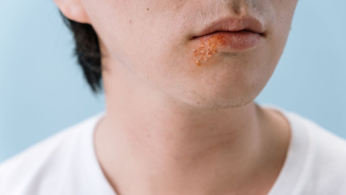
If you are currently searching online for herpes photos in different stages, you are far from alone. Finding unexpected bumps or sores can trigger immediate anxiety, and comparing what you see in the mirror to visual references online is often the very first step people take. The Herpes Simplex Virus (HSV)—whether it manifests as oral cold sores or genital lesions—follows a distinct, predictable lifecycle. Learning how these lesions evolve over time helps demystify the infection, reduces guesswork, and highlights when you are most contagious.
At the same time, visual self-diagnosis comes with real limitations. Many skin conditions mirror HSV at various points in its cycle, from ingrown hairs to contact dermatitis. While examining reliable herpes photos in different stages can help you understand the standard progression, only a clinical evaluation and lab test can give you a definitive answer.
Table of Contents
Understanding the Biology of HSV
Herpes is caused by two distinct but closely related strains: Herpes Simplex Virus Type 1 (HSV-1) and Type 2 (HSV-2). Historically, medical textbooks drew a strict line between the two, associating HSV-1 exclusively with cold sores around the mouth and HSV-2 with genital infections. Today, that boundary is much more fluid. Oral-genital contact frequently transfers HSV-1 to the genital region, making it a leading cause of initial genital outbreaks worldwide.
What makes herpes persistent is its unique relationship with the human nervous system. Once the virus enters the body through micro-tears in the skin or mucous membranes, it travels along sensory nerve pathways up to local nerve clusters, known as ganglia. There, it remains quiescent—or latent—for extended periods. Your immune system keeps the virus suppressed, but it does not clear it from the body entirely.
When triggered by factors like physical stress, illness, fatigue, intense sunlight, hormonal shifts, or mechanical friction, the virus wakes up. It travels back down the nerve fibers to the exact patch of skin served by those nerves, starting a fresh outbreak. The very first (primary) outbreak is usually the longest and most uncomfortable because the immune system has not yet developed targeted antibodies. Subsequent recurrences tend to be milder, shorter, and confined to smaller areas.
Examining Herpes Photos in Different Stages
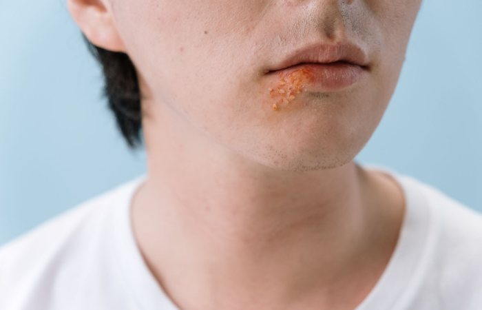
To make sense of visual references, it helps to break down an outbreak into five distinct physical phases. A typical recurrent outbreak takes anywhere from 7 to 14 days to run its full course, while a primary infection can stretch up to three weeks.
Stage 1: The Prodrome Phase (Early Warning Signals)
Before any physical bump appears on the skin, the virus announces its movement through localized sensory symptoms. As HSV reactivates and travels along nerve pathways, it irritates local nerve endings near the surface.
- Visual appearance: To the naked eye, the skin almost always looks completely normal. Near the very end of this phase, subtle localized faint redness or minor swelling might start to form, but no blisters exist yet.
- Physical sensations: You might feel a distinct tingling, itching, burning, or “electric” prickling feeling. Some individuals describe a deep, dull nerve ache radiating into the buttocks, lower back, or down the thighs.
- Contagion risk: Even though the skin looks clear, viral shedding begins during prodrome. The virus can be transmitted to a partner during this window, even without active sores present.
Stage 2: Papules (Red Bumps and Inflamed Skin)
Within 24 to 48 hours after prodromal signals begin, physical signs break through the skin surface.
- Visual appearance: Small, elevated red bumps (known medically as papules) emerge on a patch of reddened, inflamed skin. If you review clinical herpes photos in different stages, you will notice these bumps look somewhat like pimples or bug bites, but they are typically clustered together rather than spread out individually.
- Physical sensations: The area grows noticeably tender, warm to the touch, and tight. Pain becomes more localized and acute.
Stage 3: Vesicles (The Blistering Stage)
This is the signature visual phase of a herpes outbreak, where papules evolve into fluid-filled blisters.
- Visual appearance: Tightly packed clusters of small, clear, or slightly yellowish fluid-filled blisters appear on an inflamed red base. Unlike acne, which usually presents as individual blemishes, herpes vesicles gather in tight groups resembling tiny bubbles.
- Physical sensations: Discomfort peaks during this phase. The skin feels fragile, stretched, and intensely sensitive to friction or pressure.
Stage 4: Ulceration and Weeping (Open Sores)
Eventually, the thin walls of the vesicles rupture, leading to the open sore stage.
- Visual appearance: Broken blisters leave behind shallow, raw, red open sores (ulcers). On dry skin areas like the lips or outer thighs, these look like moist red craters. On mucosal surfaces—such as inside the vulva or under the foreskin—they may look like shallow, greyish-white erosions rather than distinct round blisters.
- Physical sensations: Most people report this as the most uncomfortable phase. Any contact with liquids, urine, or clothing causes sharp, stinging pain.
- Contagion risk: The liquid released from ruptured blisters contains extremely high concentrations of active virus. This is the period of highest transmissibility.
Stage 5: Crusting, Scabbing, and Skin Renewal
As your body’s immune system brings viral replication under control, the lesions transition into the final repair phase.
- Visual appearance: On dry exterior skin, shallow ulcers dry out and form honey-colored or dark brownish crusts. On moist inner mucosal tissue, scabs do not form in the traditional sense; instead, a yellowish or greyish film forms over the sore before fresh tissue builds underneath.
- Physical sensations: Pain declines noticeably, replaced by itching and tightness as the scab contracts and new pink skin grows underneath.
- Resolution: Scabs shrink and slough off naturally without leaving permanent scars, provided they are not picked at or infected by secondary bacteria.
Summary Breakdown of Outbreak Stages

Medical guides and clinical herpes photos in different stages clearly illustrate how quickly these visual cues shift over a short timeframe. The table below outlines what to expect during each phase:
| Stage Name | Typical Duration | Key Visual Characteristics | Relative Contagion Level |
|---|---|---|---|
| 1. Prodrome | 1 to 2 Days | Normal skin or faint redness; no visible blisters | Moderate (Active viral shedding) |
| 2. Papules | 1 to 2 Days | Small, elevated red bumps on inflamed skin | High |
| 3. Vesicles | 2 to 3 Days | Clustered, fluid-filled blisters on a red base | Very High |
| 4. Ulceration | 1 to 3 Days | Ruptured blisters, shallow raw open sores | Extremely High |
| 5. Crusting & Healing | 3 to 7 Days | Dry crusting, scabbing, new pink skin growth | Moderate to Low (until healed) |
Conditions That Closely Resemble Herpes
Because early bumps and shallow ulcers share features with various skin conditions, relying solely on herpes photos in different stages to diagnose yourself can lead to confusion. Several common dermatological issues can look nearly identical:
- Contact Dermatitis: Allergic reactions to laundry detergents, latex, scented soaps, or topical creams can cause localized red bumps and tiny, itchy fluid blisters.
- Folliculitis (Ingrown Hairs): Inflamed hair follicles, often caused by shaving or waxing in the pubic area, produce small red bumps with central pus pockets. Unlike herpes clusters, folliculitis centers directly around individual hair shafts.
- Canker Sores (Aphthous Ulcers): These non-contagious ulcers appear exclusively inside the mouth on soft, flexible tissues like the inner cheeks or tongue. Oral HSV-1 typically targets the firm, outer edge of the lips (the vermilion border).
- Molluscum Contagiosum: A harmless viral skin infection that produces firm, smooth, dome-shaped bumps with a characteristic central dimple. They are generally painless and do not form fluid blisters.
- Syphilis Chancres: A primary syphilis infection typically presents as a single, firm, round, and painless open sore, whereas herpes usually appears as a painful group of multiple small sores.
Getting an Accurate Medical Diagnosis
While comparing your symptoms to verified herpes photos in different stages offers useful context, a physical exam and medical testing are essential for certainty. If you notice unusual sores, scheduling a visit with a doctor or sexual health clinic is the best next step.
Diagnostic Testing Options
Healthcare providers use definitive laboratory methods to identify the virus:
- Swab Tests (PCR or Viral Culture): The gold standard for diagnosis. A provider gently swabs fluid directly from an active blister or weeping sore (Stages 3 or 4) to test for HSV DNA. PCR tests are extremely sensitive and accurate.
- Type-Specific Blood Tests: Serology tests search for IgG antibodies against HSV-1 and HSV-2. These tests are helpful if your sores have completely healed, though they indicate whether you have ever been exposed to the virus rather than proving that a specific current bump is caused by herpes.
When to Seek Immediate Medical Evaluation
Certain symptoms require prompt professional intervention:
- Lesions located near or on the eye, which can threaten vision (ocular herpes).
- High fever, severe headache, confusion, or a stiff neck accompanying an outbreak.
- Sores that become intensely hot, swollen, increasingly painful, or leak thick yellow pus, signaling a secondary bacterial infection.
- Outbreaks during pregnancy or in individuals with weakened immune systems.
Practical Care and Hygiene Strategies
Taking care of your skin during an active outbreak speeds recovery, relieves discomfort, and prevents spreading the virus to other parts of your body or to partners.
- Keep the Area Clean and Dry: Clean the affected skin gently with warm water and a mild, unscented soap. Pat the area dry carefully with a clean towel—never rub.
- Avoid Touching or Picking: Resist the urge to pop blisters or pull off scabs. Touching sores can spread the virus to other body parts (such as the eyes) or introduce staph bacteria into open wounds.
- Wear Breathable Clothing: Choose loose, soft cotton underwear and loose-fitting bottoms to reduce friction and allow airflow around genital sores.
- Practice Safe Hygiene: Wash your hands thoroughly with soap and water after any accidental contact with sores. Avoid intimate contact, kissing, or sharing towels, lip balms, or utensils from the first prodromal tingle until the skin is entirely healed.
- Manage Pain Comfortably: Applying a cool compress briefly to intact skin can dull localized pain. Over-the-counter pain relievers like ibuprofen or acetaminophen can also help manage discomfort.
Frequently Asked Questions
How long am I contagious during an outbreak?
You are considered contagious from the moment you feel the first prodromal tingling or itching until all sores have scabbed over, fallen off, and been replaced by fresh, unbroken skin.
Can I check herpes photos in different stages to tell if I have HSV-1 or HSV-2?
No. Visually, HSV-1 and HSV-2 lesions look identical under a microscope or to the naked eye. While reviewing herpes photos in different stages can show you how a typical outbreak evolves, only a type-specific PCR swab or blood test can distinguish between the two virus types.
Can you spread herpes without visible blisters?
Yes. HSV often undergoes asymptomatic viral shedding, where active virus particles travel to the skin surface without causing visible redness, bumps, or blisters. Transmission can occur during these silent periods.
Are recurrent herpes outbreaks always as severe as the first one?
Generally, no. The initial primary outbreak is almost always the most painful and widespread. Recurrent outbreaks are usually significantly milder, produce fewer blisters, heal much faster, and rarely cause systemic symptoms like fever.
Conclusion
Learning to recognize how lesions evolve—moving from an initial prodromal tingle to fluid-filled blisters, open sores, and final scabs—provides valuable clarity when dealing with a potential outbreak. While reviewing medical resources and herpes photos in different stages helps you understand what is happening on your skin, visual comparison alone cannot replace an official diagnosis. Many benign skin conditions share similar features with HSV. If you notice new or recurring sores, consult a healthcare professional for proper testing, guidance, and treatment options tailored to your needs.
Related Guides
Explore more useful resources related to this topic:
- Genetic Disorder – Introduction, Side Effects, Treatment, and More
- Understanding Neurodivergence: A Comprehensive Guide to Brain Function, Health, and Daily Well-Being
- Understanding Self Actualization: A Guide to Personal Growth and Wellbeing
- Erikson Stages of Development: A Guide to Psychosocial Growth Across Life